On global fMRI signals and simulations
Jonathan Power, Tim Laumann, Mark Plitt, Alex Martin, Steve Petersen
Trends in Cognitive Sciences
Pubmed link
Figure (.ppt)
Length limits are tight at TiCS. Here I'll substantiate one of the points we made in more detail than I could in the article, the point related to ICA not removing respiratory artifacts. ICA is good at finding spatially specific patterns in fMRI data. It's not good at finding spatially non-specific patterns, such as signals present across the entire brain. This property is baked in to ICA. If you want some evidence, images from Power, 2017, Power et al., 2017, and Carone et al., 2017 are below. Other articles like Burgess et al., 2016 or Ciric et al., 2017 further substantiate the point.
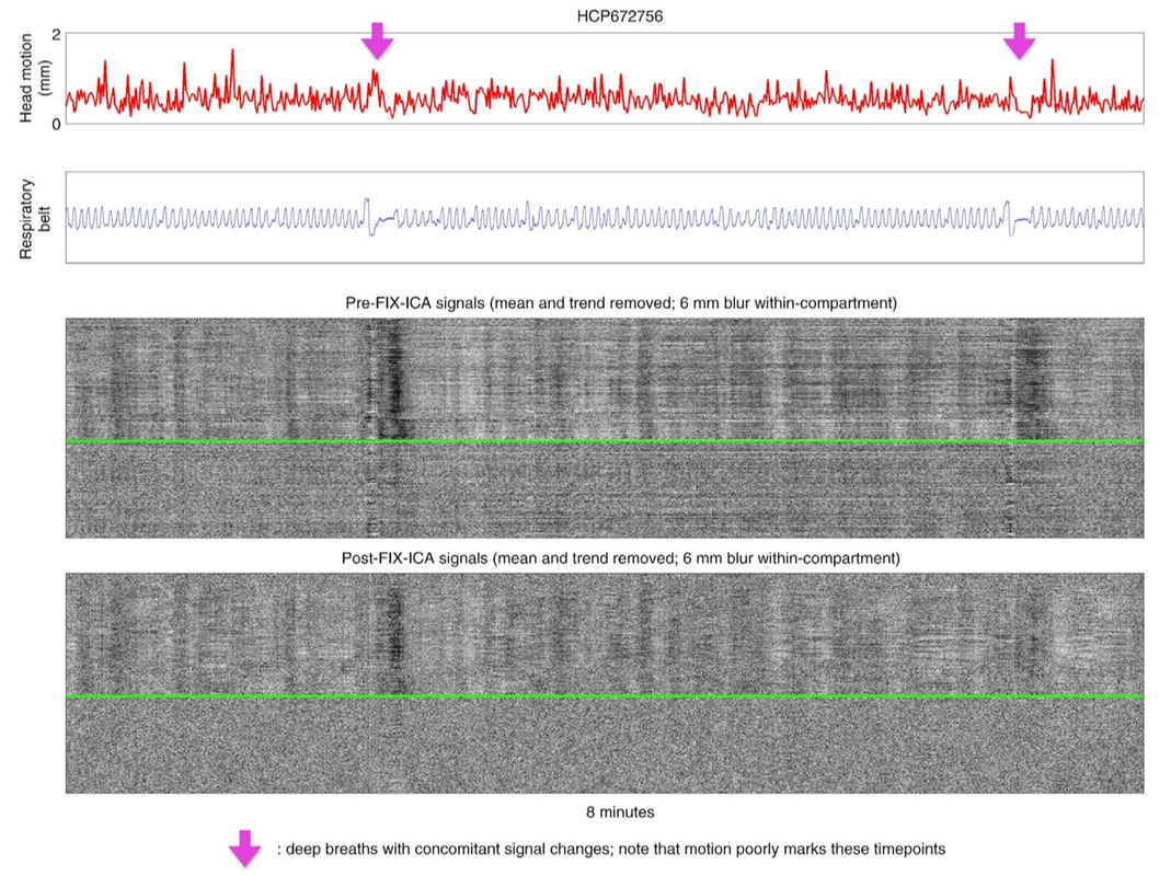
Here is a picture from my article on The Plot. The blue trace is a respiratory belt trace. The grayscale heatmap shows tens of thousands of voxel signals in gray matter (above the green line) and in white matter (below the green line). Note the two deep breaths marked by the times where the arrows are at, and then the whole-brain signal decreases (black vertical bands) that last for the next 40 seconds or so. These respiratory artifacts aren't removed by FIX-ICA.
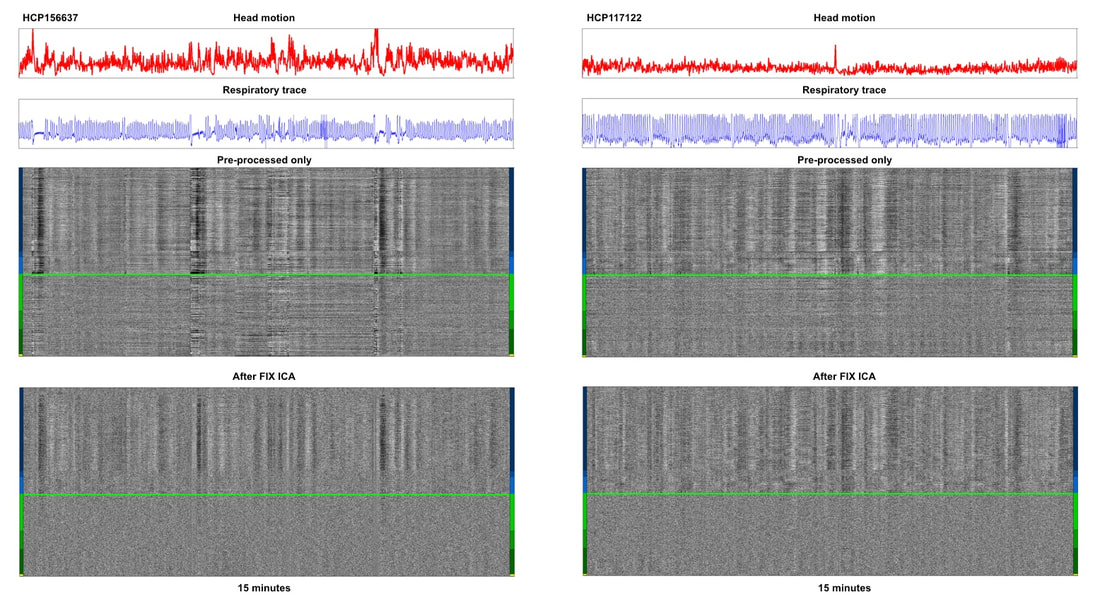
In my article on global fMRI signals (the one that was Spotlighted by TiCS), similar plots for 40 subjects of the Human Connectome Project were shown.
The two below were in a figure, you can see the rest here.
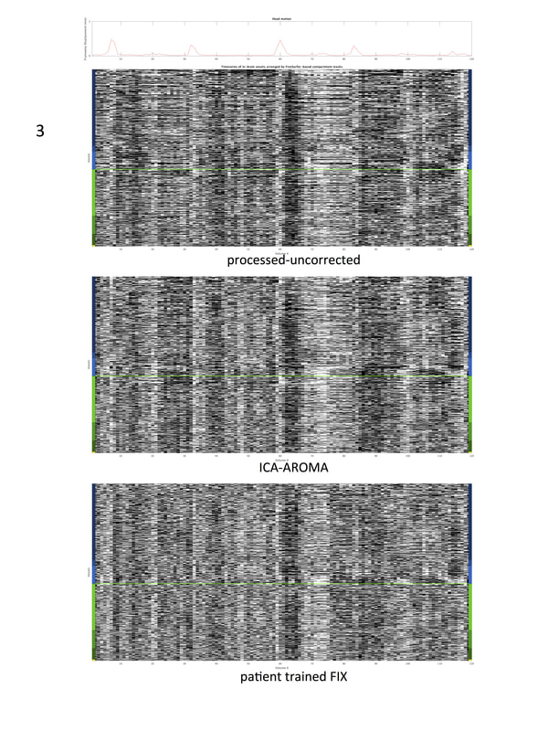
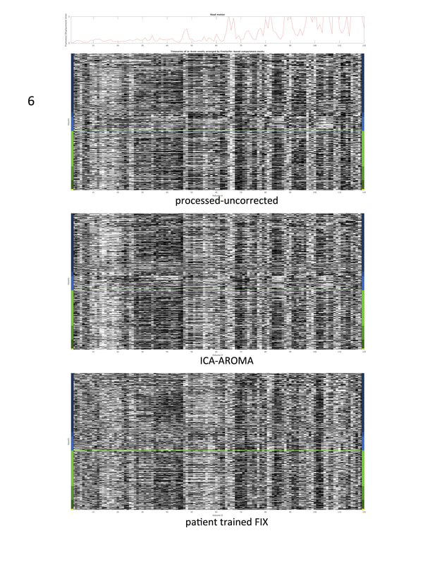
Here is evidence from Carone et al., 2017. Each of their 60 subjects underwent FIX-ICA and ICA-AROMA, and they put 20 grayplots in their supplemental materials (excerpted here). Below I put two of the plots. The configuration is as above, the red trace is motion, there is no respiratory trace shown, and gray and white matter voxels are shown with the same conventions as above.
The retention of such signals is why post-ICA timeseries are heavily dependent on indices of these signals (such as motion).


